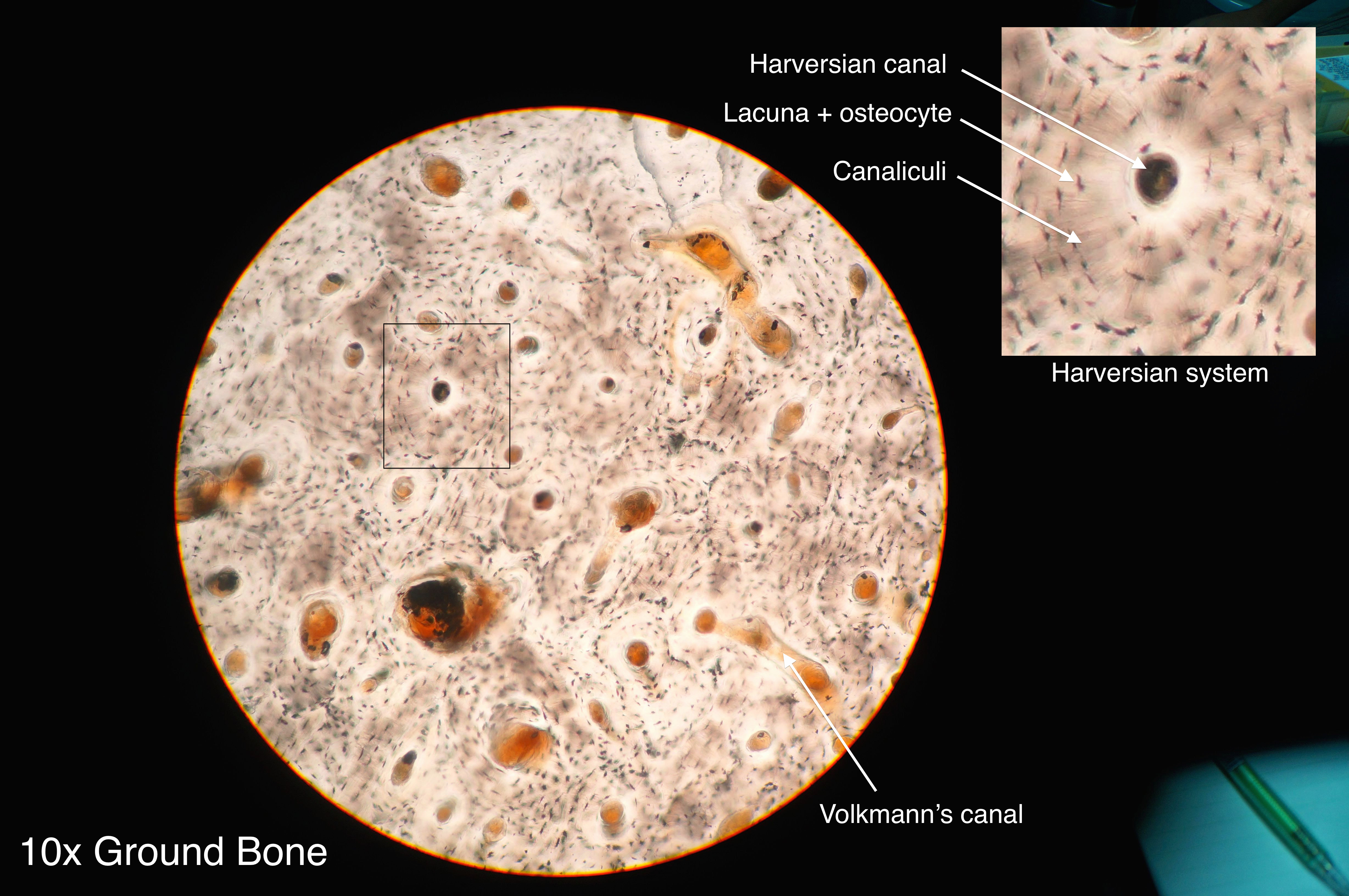
Compact Bone Diagram Microscope / Osteoblasts, Osteoclasts, Calcium, and Bone Remodeling ... / If you look at compact bone under the microscope, you will observe a highly organized arrangement of concentric circles that look like tree trunks.
Compact Bone Diagram Microscope / Osteoblasts, Osteoclasts, Calcium, and Bone Remodeling ... / If you look at compact bone under the microscope, you will observe a highly organized arrangement of concentric circles that look like tree trunks.. The scanning electron microscope (sem) is among the most frequently used instruments for examining bone. (b) in this micrograph of the osteon, you can clearly see the concentric lamellae and central canals. The collagen fibers in the more heavily stained lamellae are arranged in a circular fashion; The diagram above shows a longitudinal view of an osteon. 100x on this image you can see several of the structural units of bone tissue (osteons or haversian systems).
Bone tissue and cells under the microscope introduction. At this level of magnification, the fundamental structure of compact bone is visible. A photo taken through a microscope that shows the anatomy of compact bone with a detailed view of an osteon. Each group of concentric circles (each tree) makes up the microscopic structural unit of compact bone called an osteon (this is also called a haversian system). The cells of compact bone, which is also called cortical bone, appear to be tightly packed into a solid mass.
Fitxer:Compact bone histology 2014.jpg - Viquipèdia, l ... from upload.wikimedia.org If you look at compact bone under the microscope, you will observe a highly organized arrangement of concentric circles that look like tree trunks. Each osteon looks like a ring with a light spot in the center. Transverse section of an osteon with its haversian canal [1. 0 0000 a shoutout is a way of letting people know of a. Each group of concentric circles (each tree) makes up the microscopic structural unit of compact bone called an osteon (this is also called a haversian system). It is enveloped by lamellae in the ground substance, which may be more or less impregnated with silver nitrate. Learn vocabulary, terms, and more with flashcards, games, and other study tools. Microscopic structure of bone diagram.
In this article we will discuss about the structure of nucleus with the help of suitable diagrams.
(b) in this micrograph of the osteon, you can clearly see the concentric lamellae and central canals. Microscopic structure of bone diagram. Transverse section of an osteon with its haversian canal [1. The cells of compact bone, which is also called cortical bone, appear to be tightly packed into a solid mass. This human bone section shows the haversian canal (or osteon) structure of compact bone tissue. Each bone is a complex living organ that is made up of many cells protein fibers and minerals. Learn vocabulary, terms, and more with flashcards, games, and other study tools. Some, mostly older, compact bone is remodelled to form these haversian systems (or osteons). There are small canals that run through the bone, which allow blood vessels to penetrate it. Start studying compact bone microscopic labeling. Lamellar bone makes up the compact or cortical bone in the skeleton, such as the long bones of the legs and arms. In this article we will discuss about the structure of nucleus with the help of suitable diagrams. It can be found under the periosteum and in the diaphyses of long bones, where it provides support and protection.
Like other tissues in the body, bones are made up of specialized cells that serve different functions. It is enveloped by lamellae in the ground substance, which may be more or less impregnated with silver nitrate. The scanning electron microscope (sem) is among the most frequently used instruments for examining bone. Concentric layers of bone cells (osteocytes) and bone matrix surround the central canal. Microscopic structure of bone diagram.
Notes Ch 7 (Skeleton) from www.biologycorner.com The compact bone is the main structure in the body for support, protection, and movement. Concentric layers of bone cells (osteocytes) and bone matrix surround the central canal. The darker ring consists of layers of bone matrix made by cells called. 111 is generally a round body occupying the centre of the cell. Compact and spongy bone with dr. Under magnification you can clearly see the system of concentric circles that forms compact bone. Like other tissues in the body, bones are made up of specialized cells that serve different functions. Learn vocabulary, terms, and more with flashcards, games, and other study tools.
Like other tissues in the body, bones are made up of specialized cells that serve different functions.
At this level of magnification, the fundamental structure of compact bone is visible. 100x on this image you can see several of the structural units of bone tissue (osteons or haversian systems). Compact bone, also called cortical bone, dense bone in which the bony matrix is solidly filled with organic ground substance and inorganic salts, leaving only tiny spaces (lacunae) that contain the osteocytes, or bone cells.compact bone makes up 80 percent of the human skeleton; Each bone is a complex living organ that is made up of many cells protein fibers and minerals. The remainder is cancellous bone, which has a spongelike appearance with numerous large spaces and is found in the. Transverse section of an osteon with its haversian canal [1. The compact bone is composed of calcified extracellular material the bone matrix and 3 major cell types which are osteoblast which ssynthesize and secrete the organic components of bone matrix which include type 1 collagen fibers proteoglycans and several glycoproteins such as ostepnectin. Compact bone histology slide structure with diagram. Under magnification you can clearly see the system of concentric circles that forms compact bone. Before placing your slide on the microscope stage, remember to read the label, examine the slide with your eye and note any visible macroscopic features that might help your examination. The light spot is a canal that carries a blood vessel and a nerve fiber. It can be found under the periosteum and in the diaphyses of long bones, where it provides support and protection. This human bone section shows the haversian canal (or osteon) structure of compact bone tissue.
Learn vocabulary, terms, and more with flashcards, games, and other study tools. The diagram above shows a longitudinal view of an osteon. Learn vocabulary, terms, and more with flashcards, games, and other study tools. The remainder is cancellous bone, which has a spongelike appearance with numerous large spaces and is found in the. In three dimensions an osteon is cylindrical in shape.
Lab 1- Tissues at Texas A&M University - StudyBlue from classconnection.s3.amazonaws.com Spongy bone sharpey's fibers compact bone blood vessel periosteum perforating (volkmann's) canal blood vessel figure 7.3 microscopic structure of compact bone. The marrow in these images is red marrow. The remainder is cancellous bone, which has a spongelike appearance with numerous large spaces and is found in the. The collagen fibers in the more heavily stained lamellae are arranged in a circular fashion; The diagram above shows a longitudinal view of an osteon. There are small canals that run through the bone, which allow blood vessels to penetrate it. You can think of compact bone as being very similar. Bone tissue and cells under the microscope introduction.
Compact bone is the denser, stronger of the two types of bone tissue ( link ).
This human bone section shows the haversian canal (or osteon) structure of compact bone tissue. Transverse section of an osteon with its haversian canal [1. You can think of compact bone as being very similar. Compact bone histology slide structure with diagram. However, compact bones also serve a function in storing and releasing calcium to the. Compact bone is formed in concentric circles. The trabeculae are only a few cell layers thick. A photo taken through a microscope that shows the anatomy of compact bone with a detailed view of an osteon. Start studying compact bone microscopic labeling. In three dimensions an osteon is cylindrical in shape. Before placing your slide on the microscope stage, remember to read the label, examine the slide with your eye and note any visible macroscopic features that might help your examination. Under the microscope, bone can be divided into two types compact bone forms the outer 'shell' of bone. There are small canals that run through the bone, which allow blood vessels to penetrate it.
(b) in this micrograph of the osteon, you can clearly see the concentric lamellae and central canals compact bone diagram. (b) in this micrograph of the osteon, you can clearly see the concentric lamellae and central canals.


Posting Komentar
0 Komentar